Ever noticed a suspicious skin blister or sore? Your doctor might suggest a Tzanck test, a simple method for detecting viral skin infections. This guide provides comprehensive information about the Tzanck test—what it is, how it works, its benefits and limitations, and alternative diagnostic options. [https://www.lolaapp.com/chromesthesia] [https://www.lolaapp.com/max-msp-jitter-software]
Understanding the Tzanck Test
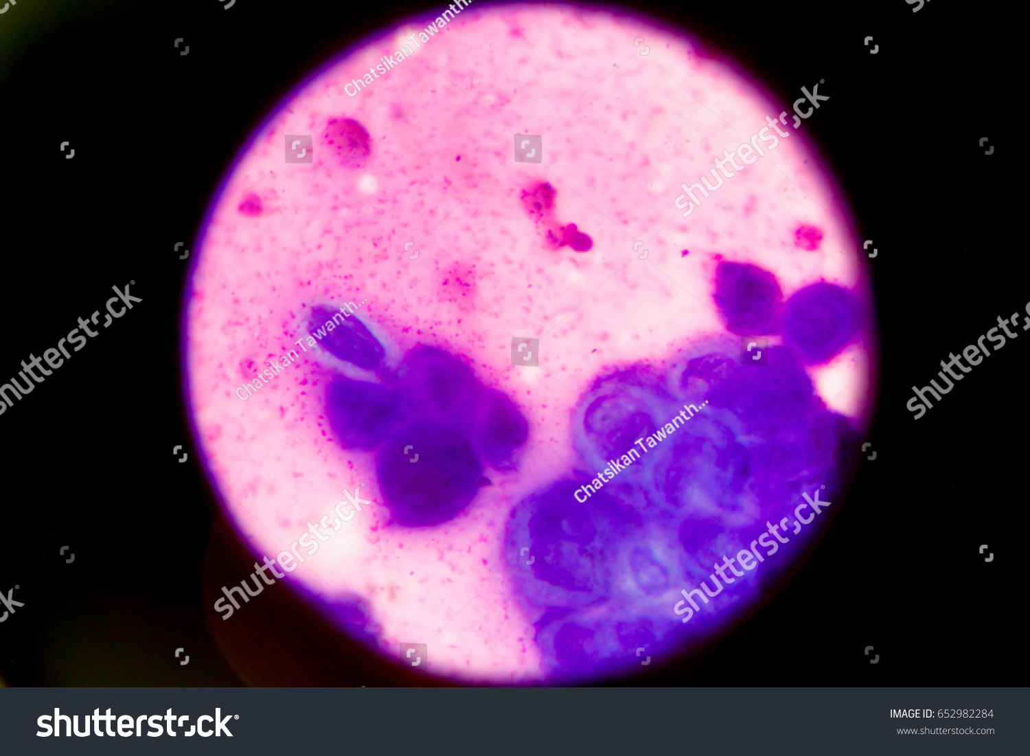
The Tzanck test, also known as the Tzanck smear, is a microscopic examination used to identify certain viral skin infections. Developed in 1947 by French dermatologist Arnault Tzanck, this test examines cells from a blister to detect the presence of viral infection.
What is the Tzanck Test Used For?
The Tzanck test helps diagnose a range of viral skin infections, including:
- Herpes Simplex Virus (HSV): This virus causes cold sores and genital herpes.
- Varicella-Zoster Virus (VZV): This virus is responsible for chickenpox and shingles.
- Pemphigus Vulgaris: This is a rare autoimmune skin disease.
How is the Tzanck Test Performed?
The Tzanck test is a relatively quick and painless procedure typically performed in a doctor’s office:
- Lesion Selection: A fresh, fluid-filled blister is selected.
- Deroofing: The top layer of the blister is carefully removed using a sterile scalpel or needle.
- Scraping: Cells are gently scraped from the blister’s base.
- Smear Preparation: The collected cells are smeared onto a glass slide.
- Fixation (Optional): The smear may be air-dried or fixed with a solution like methanol.
- Staining: A stain, such as Giemsa, Wright’s, or methylene blue, is applied to enhance cell visibility under a microscope.
- Microscopic Examination: The slide is examined under a microscope to identify specific cellular changes.
Interpreting Tzanck Test Results
Doctors look for Tzanck cells, abnormally large cells with multiple nuclei (the cell’s control center). The presence of these cells suggests a viral infection. Other cellular changes, such as acantholytic cells (detached skin cells), may also be observed.
- Positive Result: The presence of Tzanck cells likely indicates a viral infection.
- Negative Result: A negative result doesn’t completely rule out a viral infection, as false negatives can occur. Further testing may be necessary.
Advantages and Limitations of the Tzanck Test
Advantages:
- Speed: Results are typically available within minutes.
- Cost: The Tzanck test is relatively inexpensive.
- Convenience: It can be performed in a doctor’s office.
Limitations:
- Specificity: The Tzanck test cannot distinguish between different types of viral infections (e.g., HSV vs. VZV).
- Sensitivity: It can sometimes miss an infection (false negative), particularly in early stages.
- Interpretation: Accurate interpretation requires a trained healthcare professional.
Alternative Diagnostic Tests
While the Tzanck test provides valuable preliminary information, more specific tests are often needed for a definitive diagnosis:
- Viral Culture: This test involves growing the virus in a laboratory to identify it. It’s highly accurate but takes several days for results.
- PCR (Polymerase Chain Reaction): PCR detects viral DNA or RNA. It’s highly sensitive and specific, offering rapid results.
- Direct Fluorescent Antibody (DFA): This test uses fluorescent antibodies to detect specific viral antigens in the sample, providing fairly quick and specific results.
The following table compares these diagnostic methods:
| Test | Advantages | Disadvantages |
|---|---|---|
| Tzanck Test | Quick, affordable, in-office, provides preliminary info | Cannot distinguish between specific viruses, lower sensitivity |
| Viral Culture | Very accurate | Takes several days for results, needs specialized lab |
| PCR | Highly accurate, fast results | More expensive than the Tzanck test |
| DFA | Specific, fairly quick results | Requires special equipment and trained technicians |
Performing the Tzanck Test: A Step-by-Step Guide
This section offers a detailed guide for healthcare professionals on how to perform a Tzanck smear:
- Lesion Selection: Choose a fresh, fluid-filled blister (vesicle) for optimal results. Avoid crusted or older lesions.
- Deroofing: Using a sterile needle or scalpel blade, carefully remove the top layer of the blister, exposing the base.
- Scraping: Gently scrape the base of the lesion to collect cells. Avoid excessive pressure, which could cause bleeding.
- Smear Preparation: Spread the collected material thinly and evenly onto a clean glass microscope slide.
- Fixation (Optional): Air dry the smear or fix it using methanol or another appropriate fixative.
- Staining: Apply a stain, such as Giemsa, Wright’s, methylene blue, or a rapid stain like Hemacolor or Diff-Quik. Each stain has its own advantages and disadvantages in terms of sensitivity and specificity.
- Microscopic Examination: Examine the slide under a microscope, starting with a lower magnification and then increasing to higher power (oil immersion may be required). Look for multinucleated giant cells (Tzanck cells), acantholytic cells, and other characteristic changes.
Tzanck Test Stains: A Colorful Palette
Different stains can be used in the Tzanck test, each offering its own advantages:
- Giemsa: A commonly used stain, providing good overall staining but sometimes lower sensitivity.
- Methylene Blue: Research suggests methylene blue might be more sensitive than Giemsa.
- Wright’s Stain: Similar to Giemsa, with subtle differences that might be advantageous in certain situations.
- Other Stains: Less commonly used stains include Leishman stain, Papanicolaou stain, and hematoxylin and eosin (H&E).
- Rapid Stains: Hemacolor and Diff-Quik provide results in about a minute, invaluable for rapid diagnosis.
Tzanck Test for Varicella: Is It Still Relevant?
While the Tzanck test can suggest varicella (chickenpox) or shingles, it cannot definitively diagnose them. PCR testing is the gold standard for varicella diagnosis, offering superior sensitivity and specificity. However, the Tzanck test retains value in resource-limited settings or when a rapid preliminary assessment is needed.
Ongoing Research and Future Directions
Research continues to explore new diagnostic methods for viral skin infections. While the Tzanck test may not replace PCR as the gold standard, it remains a valuable tool, particularly in specific circumstances. Future research might focus on improving staining techniques and exploring the use of artificial intelligence for automated analysis of Tzanck smears.
The Tzanck test, while having limitations, offers a valuable point-of-care diagnostic tool for viral skin infections. Its speed, affordability, and ease of use make it particularly relevant in resource-constrained settings. However, confirming results with more sensitive and specific methods like PCR is important for definitive diagnosis and tailored treatment.
- Crypto Quotes’ Red Flags: Avoid Costly Mistakes - June 30, 2025
- Unlock Inspirational Crypto Quotes: Future Predictions - June 30, 2025
- Famous Bitcoin Quotes: A Deep Dive into Crypto’s History - June 30, 2025


