Unveiling the hidden wonders of the world unseen, this article takes you on a captivating journey into the realm of microscopy. Get ready to embark on an exploration of different types of microscopes, the extraordinary tools that reveal the microscopic universe in all its intricate glory. From the conventional light microscope to cutting-edge imaging technologies, we will dive deep into the fascinating world of microscopy, shedding light on its various techniques and their remarkable applications. So, buckle up and prepare to see the minuscule in a whole new light, as we unravel the secrets of these remarkable scientific marvels.

Types Of Microscope
Microscopes have revolutionized our understanding of the world by allowing us to explore the unseen. There are various types of microscopes, each with its unique capabilities and applications. In this article, we will delve into the intricacies of different types of microscopes, shedding light on their functions and revealing the remarkable advancements in microscopy.
Compound Microscopes
One of the most common types of microscopes is the compound microscope. It utilizes lenses and light to illuminate a specimen, allowing for image-gathering. These microscopes are widely used in laboratories, educational settings, and research facilities. The compound microscope magnifies the specimen by passing light through the objective lens and then through the eyepiece to create a magnified image.
“Compound microscopes are the workhorse of microscopy, offering a versatile tool for a wide range of scientific inquiries and educational purposes.”
Confocal Microscopes
Advancements in microscopy have given rise to specialized tools like confocal microscopes. These instruments use lasers to scan a specimen and create high-resolution images with depth-selection. By pinpointing the focal plane and excluding out-of-focus light, confocal microscopes can produce sharp images with exceptional clarity. These microscopes are particularly useful in studying biological samples, providing detailed insights into cell structures and dynamics.
“Confocal microscopes unlock the hidden details of specimens, revealing their intricate beauty with astonishing precision.”
Stereoscopic Microscopes
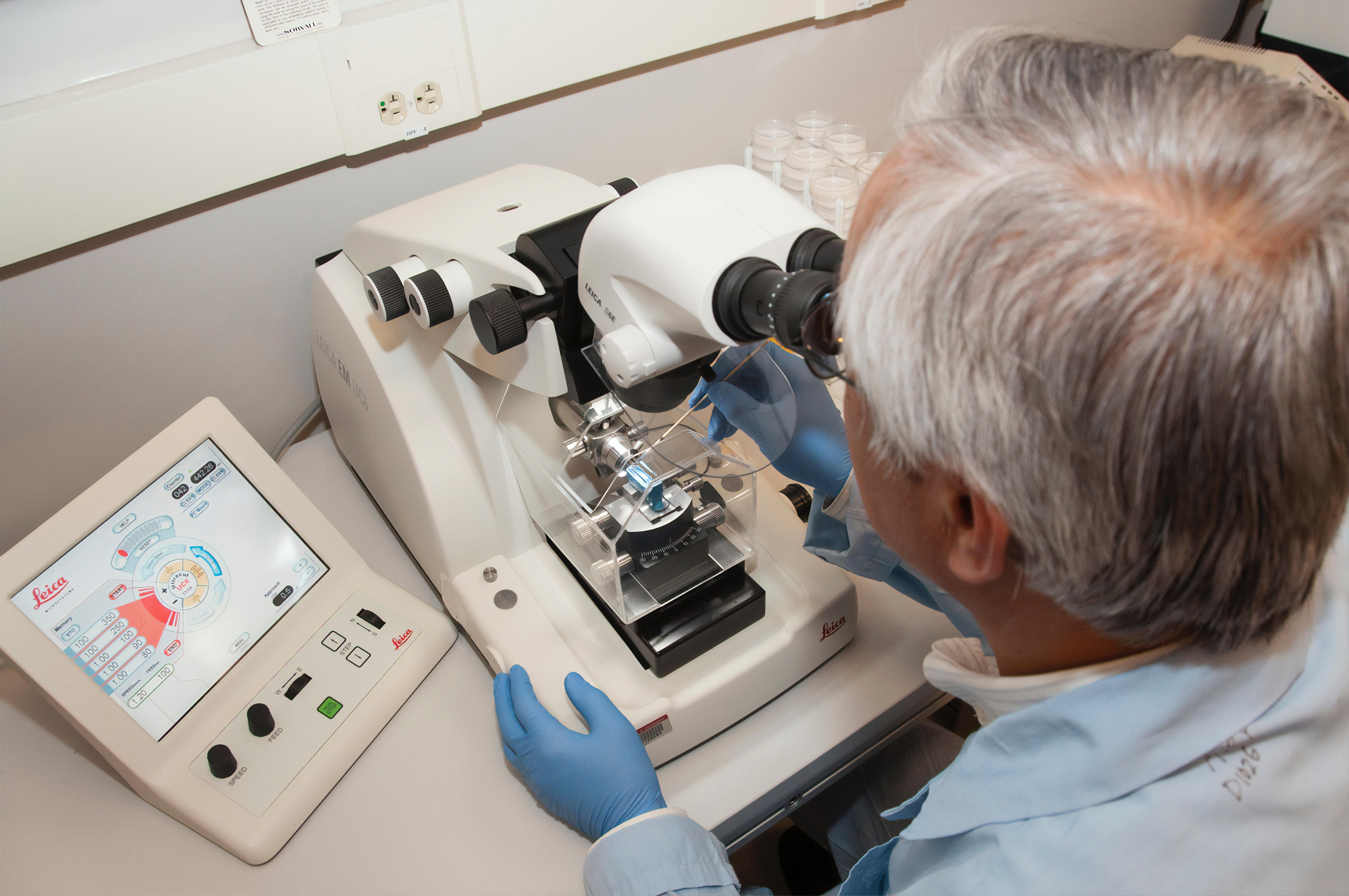
In labs and educational settings, you will often find stereoscopic microscopes in action. These microscopes employ a top light source to illuminate the specimen from above, allowing for three-dimensional observation. This design enables users to examine larger objects or samples with intricate structures. Stereoscopic microscopes are indispensable tools for various fields, including forensics, quality control, and dissection.
“Stereoscopic microscopes bring the microscopic world to life, unmasking the hidden wonders of larger specimens.”
Electron Microscopes
When it comes to achieving the highest power and resolution, electron microscopes take the lead. Instead of using light, these microscopes employ accelerated electrons to create a digital image. By utilizing the short wavelength of electrons, electron microscopes overcome the limitations of traditional optics, revealing even the tiniest details of samples. Electron microscopes play a vital role in fields such as materials science, nanotechnology, and biomedical research.
“Electron microscopes peer into the atomic realm, unraveling the mysteries hidden within the tiniest building blocks of our world.”
Simple Microscopes
In contrast to compound microscopes, simple microscopes use a single lens to magnify and view small objects. While they may not possess the same complexity or magnification power as other types of microscopes, simple microscopes are valuable tools in certain applications. They offer a straightforward and accessible way to examine objects, making them ideal for educational purposes and fieldwork.
“Simple microscopes provide a clear window into the microcosmos, enabling us to observe the minute features that surround us.”
Phase Contrast Microscopes
Phase contrast microscopes employ optical techniques to enhance the contrast of transparent samples. They enable scientists to observe and analyze structures that would otherwise be difficult to see with conventional microscopy techniques. Phase contrast microscopy has revolutionized the study of living cells, allowing for non-invasive imaging and detailed examination of cellular processes.
“Phase contrast microscopes reveal the invisible, unlocking the world of transparent specimens with unparalleled clarity.”
Fluorescence Microscopes
In certain studies, researchers need to label specific structures within a specimen. Fluorescence microscopes come to the rescue by utilizing fluorescent dyes that attach to target structures, making them visible under specific wavelengths of light. This technique is widely used in fields such as cell biology, immunology, and neuroscience to investigate the localization and dynamics of specific molecules within cells and tissues.
“Fluorescence microscopes illuminate the hidden actors within cells, allowing us to witness their intricate dance in living color.”
Scanning Probe Microscopes
When it comes to investigating surfaces and structures at the atomic level, scanning probe microscopes take center stage. These instruments use a probe to scan a sample’s surface, capturing fine details with exceptional precision. Scanning probe microscopes have made significant contributions to nanotechnology, material science, and surface characterization, enabling researchers to explore the world at a breathtakingly small scale.
“Scanning probe microscopes unveil the atomic landscapes, guiding us through the tiny terrains that shape our material world.”
Low-Powered and High-Powered Microscopes
Microscopes come in different power ranges, suited for different purposes. Low-powered microscopes are designed to observe larger, more visible features and are commonly used in educational settings. They provide opportunities for students and enthusiasts to explore the world unseen on a macroscopic level.
On the other hand, high-powered microscopes are built for resolving smaller features and offer greater magnification capabilities. These microscopes are indispensable in scientific research, where intricate details and minute features need to be examined.
“Low-powered and high-powered microscopes each serve a unique purpose, offering windows into the world at different levels of magnification.”
Optical Microscopes
Finally, we come full circle to the most familiar type of microscope: the optical microscope. These microscopes use glass lenses to form an image and are the foundation of many other types of microscopes. Optical microscopes are versatile tools that have paved the way for countless scientific breakthroughs, allowing researchers to explore the microscopic world with ease.
“Optical microscopes remain the backbone of microscopy, offering a glimpse into the hidden wonders that lie within our reach.”
In conclusion, the world of microscopy is vast and diverse, with each type of microscope offering unique capabilities and applications. From the compound microscope to the confocal microscope, the stereoscopic microscope to the electron microscope, each instrument has expanded our understanding of the microscopic realm. By illuminating the invisible and magnifying the minute, microscopes have unlocked wonders that were once beyond our imagination. So, let us embark on this extraordinary journey through the unseen, exploring the world of microscopes and unraveling the mysteries of the microscopic domain.
Microscopes have been revolutionizing our understanding of the world around us for centuries. If you’re fascinated by the incredible capabilities of these scientific instruments, you’ll definitely want to check out these mind-blowing facts about the microscope. Prepare to be amazed as you dive into the microscopic realm and discover a whole new world of wonders. Click here to unveil the captivating secrets of the microscope: facts about the microscope.
Types Of Microscope
A fascinating world lies beyond the naked eye, waiting to be explored. Discover the wonders of science with different types of microscopes. These incredible instruments unveil the hidden realms, allowing us to zoom into the smallest details of life. Delve into the microscopic universe with ease by exploring the various types of microscopes available. From compound microscopes to electron microscopes, each offers a unique lens through which we can witness the intricate beauty of cells, organisms, and beyond. Embark on a journey of knowledge and curiosity, and click here to explore different types of microscopes. And for a comprehensive understanding of the subject, dive into the realm of science with various types of microscopes here.
Types of Microscopes and Their Functions
[youtube v=”nuJZtXohFFQ”]
Overview
Microscopes are essential tools in laboratories for examining objects that are too small to be seen with the naked eye. There are various types of microscopes, each with its own working principle and application. In this article, we will explore the different types of microscopes and their functions.
Simple Microscope
The simple microscope, invented by Anthony van Leeuwenhoek in the 17th century, was the first microscope ever created. It consists of a single lens that magnifies an object through angular magnification. Simple microscopes, such as magnifying glasses, loops, and eyepieces, provide an erected, enlarged virtual image to the viewer. They are commonly used in education and fieldwork.
“Simple microscopes use a single lens to enlarge objects, making them valuable for educational purposes and fieldwork.”
Compound Microscope
The compound microscope is a laboratory instrument with high magnification power, consisting of multiple lenses. It is widely used for studying the structural details of cells, tissues, and organs. Compound microscopes can magnify tiny objects up to a thousand times. They typically have an objective lens for magnification and an eyepiece lens for viewing.
“Compound microscopes are the most common type of microscope used for a wide range of scientific and educational purposes.”
Light Microscope
Light microscopes use visible light and magnifying lenses to examine small objects in finer detail than the naked eye allows. They are classified into four categories: bright field microscope, dark field microscope, phase contrast microscope, and fluorescent microscope.
Bright Field Microscope: In a bright field microscope, the specimen appears dark against a bright background. It is commonly used to study the outer structure of microorganisms.
Dark Field Microscope: In a dark field microscope, the specimen appears bright against a dark background. This microscope is used to distinguish unstained, thin, living cells that are not visible under a simple microscope.
Phase Contrast Microscope: Phase contrast microscopes create contrast differences between cells and water, allowing the visualization of unpigmented living cells that are not visible in a light microscope. They are used for studying the shape and motility of microorganisms.
Fluorescent Microscope: Fluorescent microscopes use fluorescent dyes to stain specimens, which are then exposed to ultraviolet rays. This microscope is particularly useful in medical laboratories for identifying pathogens and specific proteins.
“Light microscopes use visible light and magnifying lenses to examine small objects in finer detail. They are classified into four categories: bright field, dark field, phase contrast, and fluorescent microscopes.”
Electron Microscope
Electron microscopes use electrons instead of light to create high-resolution images with exceptional clarity. They are widely used in fields such as materials science, nanotechnology, and biomedical research. There are three types of electron microscopes: scanning electron microscope, transmission electron microscope, and confocal microscope.
Scanning Electron Microscope (SEM): SEM produces an image of a specimen by scanning the surface with a focused beam of electrons. It is used to study the surface area of microorganisms in detail.
Transmission Electron Microscope (TEM): TEM passes an electron beam through a specimen to form an image. It is used to study the internal structure of a specimen.
Confocal Microscope: Confocal microscopy is an optical imaging technique that uses a spatial pinhole to increase the optical resolution and contrast of a micrograph. It captures multiple two-dimensional images at different depths within an object, enabling the reconstruction of three-dimensional structures.
“Electron microscopes use electrons as a source of illumination and provide high-resolution images. There are three types of electron microscopes: scanning, transmission, and confocal microscopes.”
Conclusion
Microscopes play a crucial role in various scientific and educational fields. Understanding the different types of microscopes and their functions can help researchers and scientists choose the most suitable tool for their specific needs. Whether it’s simple microscopes for educational purposes or electron microscopes for advanced research, each type of microscope contributes to our understanding of the microscopic world.
“Microscopes are invaluable tools in scientific research, enabling the exploration of the microscopic world and enhancing our understanding of various fields of study.”
FAQ
Question 1
What are the different types of microscopes commonly used in scientific research?
Answer 1
There are several types of microscopes used in scientific research, including compound microscopes, confocal microscopes, stereoscopic microscopes, electron microscopes, simple microscopes, phase contrast microscopes, fluorescence microscopes, scanning probe microscopes, low-powered microscopes, high-powered microscopes, and optical microscopes.
Question 2
How does a compound microscope work?
Answer 2
A compound microscope uses lenses and light to illuminate a specimen for image-gathering. The specimen is placed on a slide and viewed under the microscope’s lenses, which magnify the image. Light passes through the slide and lenses, allowing the viewer to observe the specimen in detail.
Question 3
What is the function of a confocal microscope?
Answer 3
Confocal microscopes use lasers to scan a specimen and create high-resolution images with depth-selection. These microscopes are especially useful for studying three-dimensional structures and can provide detailed information about the depth and location of specific components within a sample.
Question 4
What is the difference between a low-powered microscope and a high-powered microscope?
Answer 4
Low-powered microscopes are used to observe larger, more visible features, while high-powered microscopes are used to resolve smaller features. High-powered microscopes have greater magnification capabilities and can provide more detailed images of specimens.
Question 5
What are the advantages of using an electron microscope?
Answer 5
Electron microscopes use accelerated electrons instead of light to create a digital image and have the highest power. They can provide extremely high-resolution images and allow scientists to observe structures at the atomic level. Electron microscopes are especially useful for studying materials that are too small or transparent to be observed with other types of microscopes.
- Mastering Leader in Spanish: The Complete Guide - April 19, 2025
- Uncovering Surprising Parallels: England Size Compared to US States - April 19, 2025
- Old Mexico Map: Border Shifts 1821-1857 - April 19, 2025


