Nephrons are the microscopic workhorses of the kidneys, responsible for filtering blood and producing urine. Understanding their structure and function is crucial for grasping kidney physiology and diagnosing related diseases. This comprehensive guide will delve into nephron labeling, exploring each component’s role in maintaining the body’s delicate balance. We’ll also provide interactive resources, like quizzes and diagrams, to enhance your learning experience.
Decoding the Nephron: Structure and Function
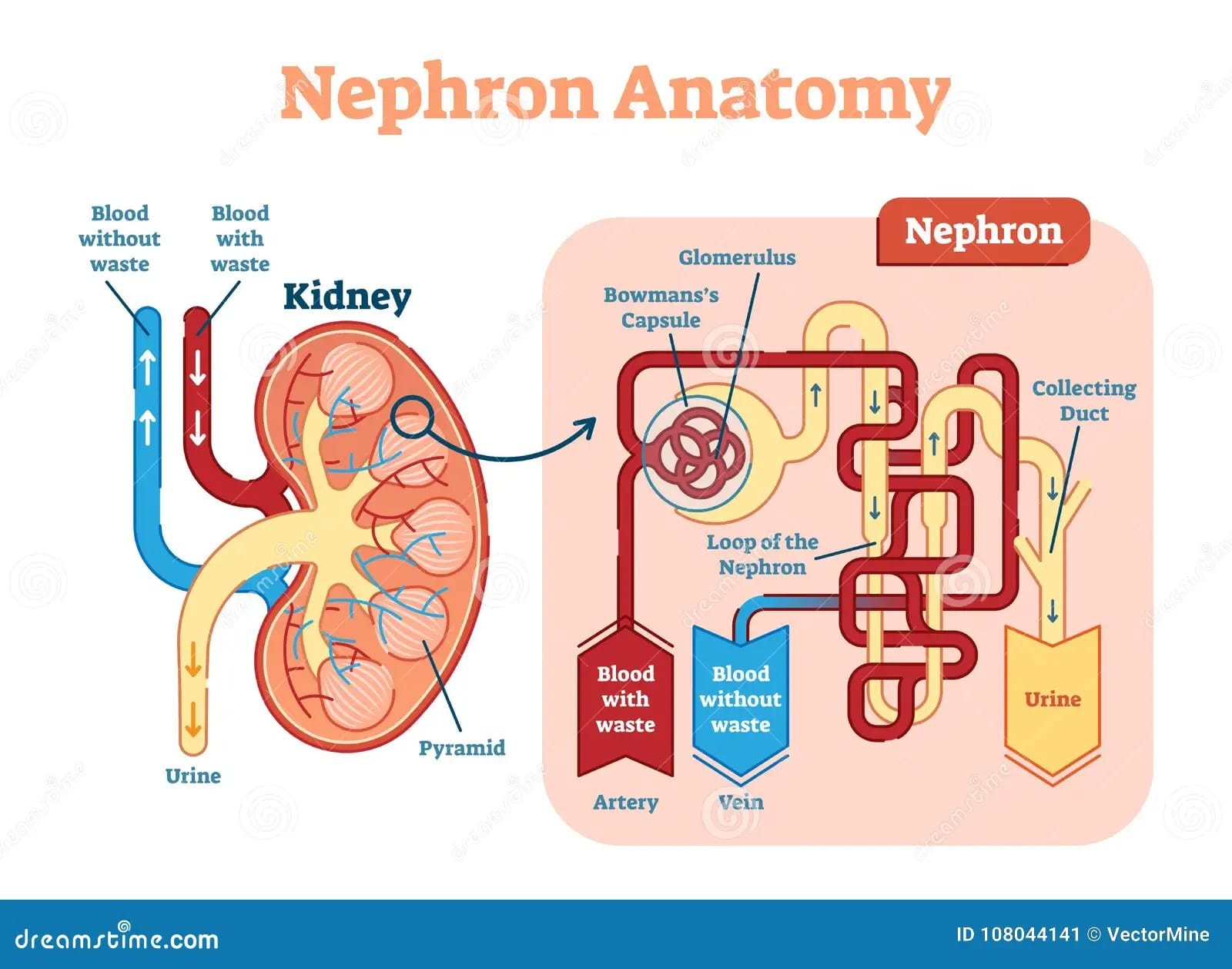
Each nephron, a self-contained filtering unit, consists of two main parts: the renal corpuscle and the renal tubule. Visualizing these structures is key to understanding their intricate interplay.
The Renal Corpuscle: The Filtration Starting Point
Imagine the renal corpuscle as the initial filtering station. It houses the glomerulus, a network of capillaries, encased by Bowman’s capsule. Blood pressure forces fluid and small molecules from the glomerulus into Bowman’s capsule, forming the initial filtrate. This filtrate contains both waste and essential substances that the body needs to reclaim.
The Renal Tubule: Refining the Filtrate
From Bowman’s capsule, the filtrate enters the renal tubule, a long, winding tube divided into distinct sections:
- Proximal Convoluted Tubule (PCT): This twisted section reabsorbs essential substances like water, glucose, amino acids, and salts back into the bloodstream.
- Loop of Henle: This hairpin-shaped loop dips into the medulla of the kidney. Its descending limb allows water reabsorption, while the ascending limb reabsorbs salts, contributing to urine concentration.
- Distal Convoluted Tubule (DCT): The DCT fine-tunes the balance of electrolytes and water, further refining the filtrate. Hormones like aldosterone and antidiuretic hormone (ADH) act on this segment to regulate electrolyte and water balance.
- Collecting Duct: This final channel collects urine from multiple nephrons and carries it towards the renal pelvis and ultimately, the bladder.
Nephron Types: Cortical vs. Juxtamedullary
Two main types of nephrons exist, distinguished by their location and Loop of Henle length:
- Cortical Nephrons: Located primarily in the cortex, these nephrons have shorter Loops of Henle and are most numerous. They perform the bulk of filtration and reabsorption.
- Juxtamedullary Nephrons: These nephrons have longer Loops of Henle that extend deep into the medulla. They play a crucial role in concentrating urine, conserving water, and maintaining osmotic balance.
Nephron Histology: A Microscopic View
Each nephron segment has a unique cellular makeup tailored to its specific function. For instance, the PCT has a brush border of microvilli to increase surface area for reabsorption. The thin segments of the Loop of Henle have flattened cells suited for passive transport, while the thick ascending limb has thicker cells involved in active transport of salts.
Nephron Function: Filtration, Reabsorption, and Secretion
Nephrons perform three essential functions:
- Filtration: Blood is filtered in the renal corpuscle, forming the initial filtrate.
- Reabsorption: Essential substances are reabsorbed from the filtrate back into the bloodstream throughout the renal tubule.
- Secretion: Waste products and excess ions are actively secreted into the tubule, adding to the urine.
These processes work together to maintain homeostasis by regulating:
- Blood Volume and Pressure: Nephrons control water and salt balance, influencing blood volume and therefore, blood pressure.
- Electrolyte Balance: Reabsorption and secretion of electrolytes like sodium, potassium, and chloride maintain their optimal concentrations in the blood.
- Waste Removal: Nephrons filter and excrete metabolic waste products such as urea and creatinine.
- Acid-Base Balance: Nephrons play a role in regulating blood pH by controlling the excretion of hydrogen ions and bicarbonate.
Nephron Labeling Techniques: Unveiling the Microscopic World
Scientists use various techniques to study and label different parts of the nephron, providing insights into kidney function and disease:
- Molecular Markers: These “tags” attach to specific proteins within the nephron, allowing scientists to differentiate between segments.
- Immunohistochemistry: Antibodies tagged with fluorescent dyes or enzymes visualize specific proteins within nephron cells, revealing their precise location.
- In Situ Hybridization: Labeled RNA or DNA probes detect specific gene sequences within nephron cells, offering information about gene activity.
These techniques, combined with advanced microscopy like fluorescence and confocal microscopy, enable detailed visualization and analysis of nephrons, even observing activity in real time.
The Clinical Relevance of Nephron Labeling
Nephron labeling has far-reaching implications in clinical practice:
- Disease Diagnosis: Changes in the expression of molecular markers can be early warning signs of kidney disease, aiding diagnosis and guiding treatment.
- Treatment Development: Studying labeled nephrons helps researchers investigate disease progression and develop new therapies.
- Nephron Number Assessment: Determining nephron number is important for overall kidney health assessment. Traditional clinicopathological assessment relies on post-mortem kidney biopsies. However, promising in vivo techniques are emerging that use non-invasive imaging methods to estimate nephron number in living patients. These methods may revolutionize early detection and monitoring of kidney disease. Dive into the fascinating world of lactose fermenting gram negative rods, which can sometimes be implicated in urinary tract infections impacting nephron function.
Interactive Nephron Labeling Resources: Engaging with the Microscopic World
Reinforce your understanding of nephron anatomy and function with these interactive resources:
- Interactive Labeling Activities: Drag-and-drop exercises provide hands-on learning and instant feedback.
- Quizzes and Worksheets: Test your knowledge and identify areas needing further study.
- 3D Nephron Models: Explore nephron structure in a dynamic, immersive way. Many models also include animations showing fluid and electrolyte movement.
Ongoing Research and Future Directions
Our understanding of nephrons is constantly evolving. Ongoing research is exploring several areas:
- Nephron Dysfunction in Disease: Researchers are investigating how specific diseases impact nephron function and exploring potential therapeutic targets.
- Regenerative Medicine: Some studies suggest the possibility of regenerating damaged nephrons, which could revolutionize kidney disease treatment.
- Comparative Nephron Anatomy: Studying nephron adaptations across different species provides insights into evolutionary strategies for maintaining fluid and electrolyte balance.
This guide offers a foundational understanding of nephrons and their vital role in maintaining health. While current knowledge provides a comprehensive framework, ongoing research continues to refine our understanding and unlock new avenues for diagnosis, treatment, and potentially even prevention of kidney disease. Embrace the interactive resources and delve deeper into the fascinating world of nephrons!


