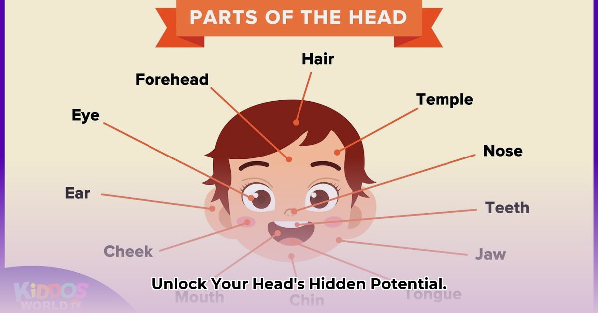
The Intricate Architecture of Your Head: Cranium, Face, and Their Dynamic Interplay
The human head, a marvel of biological engineering, houses the command center of our being – the brain. This complex structure is not merely a collection of bones and muscles but a dynamic system enabling us to perceive, interact with, and interpret the world around us. This guide delves into the key components of this system, exploring the cranium, facial bones, muscles, and the ongoing research that continues to unravel its mysteries. For a visual aid, check out this helpful 3D muscle model.
The Cranium: The Brain’s Fortress
The cranium, a robust bony shell, provides the ultimate protection for the delicate brain tissue within. Comprised of eight intricately joined bones, this structure forms a formidable barrier against external forces.
Frontal Bone: Shapes the forehead and contributes to the upper portion of the eye sockets, playing a crucial role in facial structure and protecting the frontal lobe of the brain.
Parietal Bones (2): These paired bones form the roof and upper sides of the cranium, providing crucial protection to the parietal lobes of the brain. Their inner surfaces feature grooves for blood vessels supplying the brain.
Temporal Bones (2): Located on the sides of the skull, these bones house the delicate structures of the middle and inner ear, essential for hearing and balance. They also contribute to the temporomandibular joint, enabling jaw movement. The mastoid process, a bony prominence behind the ear, serves as an attachment point for neck muscles.
Occipital Bone: Situated at the back and base of the skull, the occipital bone protects the occipital lobe of the brain and contains the foramen magnum, a vital opening through which the spinal cord connects to the brainstem.
Sphenoid Bone: A complex, butterfly-shaped bone at the base of the skull, the sphenoid bone forms part of the eye sockets and the floor of the cranial cavity. It also houses the pituitary gland, a master gland regulating numerous bodily functions.
Ethmoid Bone: Located between the eyes, the ethmoid bone contributes to the nasal cavity and the orbits. It also plays a role in olfaction, as the olfactory nerves pass through it to reach the brain.
These cranial bones are joined by fibrous joints called sutures, allowing for growth during childhood and adolescence. The intricate interlocking of these sutures contributes to the structural integrity of the cranium.
The Facial Skeleton: Expression, Senses, and More
Beyond its aesthetic contribution, the facial skeleton plays a critical role in supporting sensory organs, facilitating speech, and enabling a wide range of facial expressions. Fourteen bones contribute to the intricate framework of the face.
Maxilla (2): These paired bones form the upper jaw, supporting the upper teeth and contributing to the hard palate, nasal cavity, and eye sockets.
Mandible: The only movable bone in the skull, the mandible forms the lower jaw, crucial for chewing, speaking, and facial expressions.
Zygomatic Bones (2): Commonly known as the cheekbones, these bones contribute to the prominence of the cheeks and form part of the lateral wall of the eye sockets.
Nasal Bones (2): These small bones form the bridge of the nose, contributing to its shape and supporting the nasal cartilage.
Lacrimal Bones (2): Situated at the inner corner of each eye socket, these tiny bones contain the nasolacrimal ducts, which drain tears into the nasal cavity.
Palatine Bones (2): These L-shaped bones form the posterior portion of the hard palate and part of the nasal cavity and eye sockets.
Vomer: This thin, flat bone forms the lower part of the nasal septum, dividing the nasal cavity into two halves.
Inferior Nasal Conchae (2): These curved bones within the nasal cavity increase the surface area, helping to warm and humidify inhaled air.
The complex interplay of these bones allows for the subtle nuances of human facial expression, from a slight smile to a furrowed brow.
Muscles: The Engines of Expression and Mastication
The muscles of the head are divided into two main groups: the muscles of facial expression and the muscles of mastication.
Muscles of Facial Expression: These numerous small muscles, located just beneath the skin, control facial movements, enabling a vast repertoire of expressions. These muscles include the orbicularis oculi (closes the eyelids), orbicularis oris (controls mouth movements), and zygomaticus major (lifts the corners of the mouth in a smile).
Muscles of Mastication: Primarily responsible for chewing, these powerful muscles include the temporalis (elevates the mandible), masseter (closes the jaw), and medial and lateral pterygoids (move the mandible sideways and forward).
Unraveling the Mysteries: Ongoing Research and Clinical Significance
The intricate structures of the head continue to be a subject of ongoing research. Scientists are exploring the complex interplay of nerves, blood vessels, and lymphatic systems within the head, seeking to understand how they contribute to sensory perception, motor control, and overall head and neck function. Advanced imaging techniques, such as MRI and CT scans, provide invaluable tools for visualizing these structures, aiding in diagnosis and treatment planning. This knowledge is particularly critical in fields like neurosurgery, where precise anatomical understanding is essential for successful surgical interventions. Further research is also focused on age-related changes in head and neck anatomy, seeking to develop targeted therapies for conditions like temporomandibular joint disorders and age-related hearing loss. The future holds exciting possibilities for personalized medicine, tailoring treatments to individual anatomical variations and maximizing patient outcomes.
Hello, My love for writing is rivaled only by my passion for in-depth research. While many may skim the surface, I dive deep, seeking the hidden gems of information that give a story texture and depth. Every word I write is backed by hours of exploration, ensuring that my work is not just compelling, but also meticulously accurate. Join me on a journey where every piece is a deep dive into the realms of knowledge and storytelling.