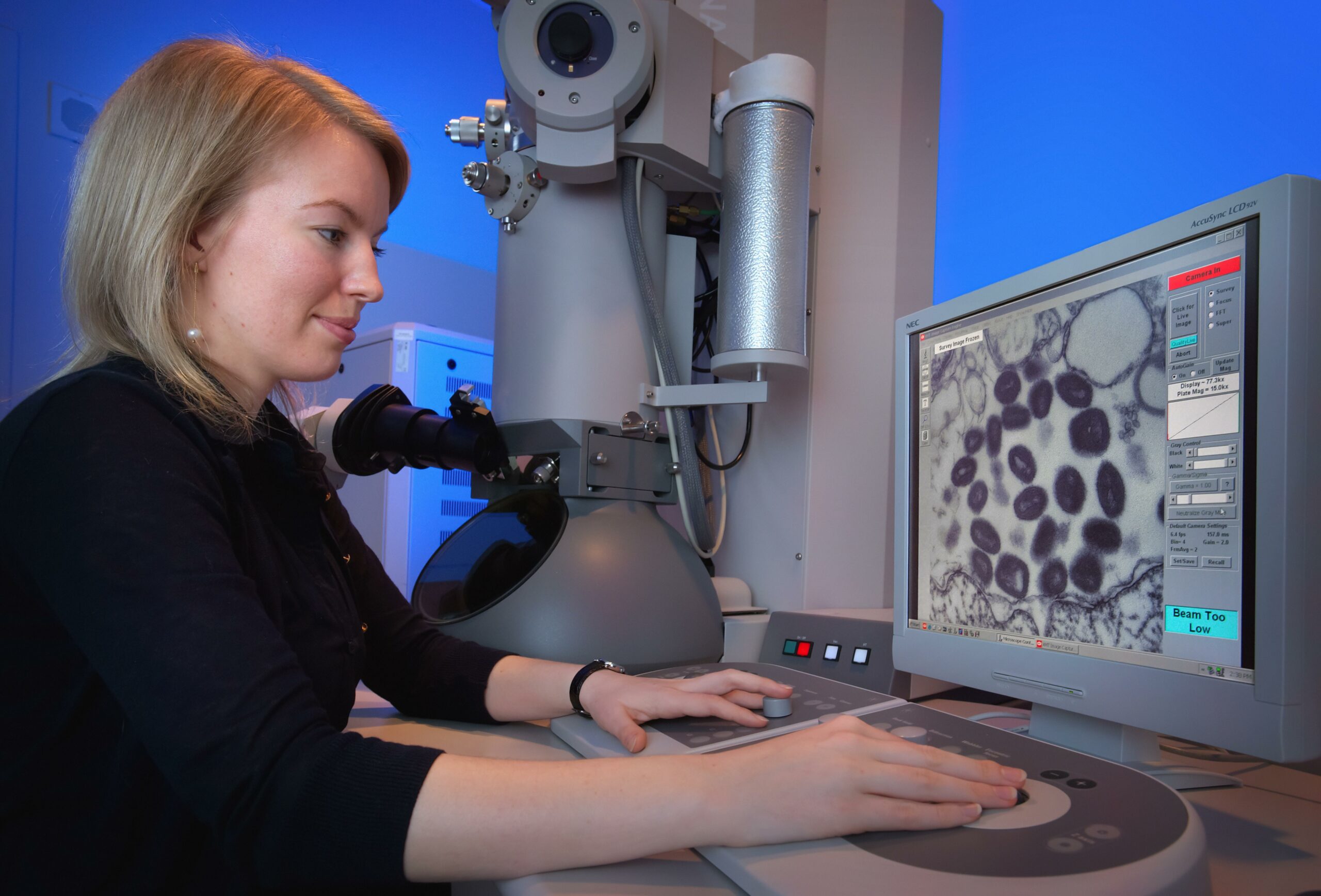
Prepare to embark on a fascinating journey into the intricate world of microscopes, where the unimaginable becomes visible, and the miniaturized becomes grandeur. In this article, we will unravel the astonishing capabilities and inherent limitations of these remarkable tools that have revolutionized the way we explore the scientific frontier. As we delve into the microscopic realm, we will witness the breathtaking wonders that lie beyond the naked eye’s gaze. Brace yourself for an exploration of the unseen, where curiosity meets scientific prowess, and the unseen becomes the seen. Welcome to the awe-inspiring world of microscopes, where the boundaries of discovery are constantly pushed and the smallest details unlock profound mysteries.

Astonishing Capabilities and Limitations of Microscopes
Microscopes have revolutionized the world of scientific discovery, allowing us to explore the mysteries of the microscopic realm with unprecedented clarity and detail. From the astonishing capabilities to the inherent limitations, these powerful tools have transformed our understanding of the microscopic world. In this article, we will delve into the remarkable capabilities and limitations of microscopes, shedding light on their incredible potential and the boundaries they encounter along the way.
The Power of Light Microscopes
Light microscopes serve as the workhorses of biological research, enabling scientists to observe cells, tissues, and even living organisms in great detail. Their high resolution makes them invaluable tools for studying the intricate structures of the microscopic world. With the ability to magnify specimens up to thousands of times, light microscopes open new avenues for unraveling the complexities of life.
But like any tool, light microscopes have their limitations. The principal limitation lies in their resolving power, which determines the smallest detail that can be discerned. Despite impressive advancements, light microscopes are still bound by the laws of physics, meaning there is a boundary beyond which finer details become blurred and indistinguishable.
“The resolving power of light microscopes, while impressive, has inherent limitations in capturing the smallest structures within a specimen.”
Unveiling Surfaces with Electron Microscopes
When it comes to viewing surface details at the nanoscale, electron microscopes take center stage. These powerful instruments utilize a beam of electrons instead of light, enabling much higher resolution than their optical counterparts. By harnessing the short wavelengths of electrons, electron microscopes can reveal intricate surface features with astonishing clarity.
However, electron microscopes also come with their own set of limitations. One significant limitation is their dependence on false colorization to produce images. Unlike light microscopes, which capture color naturally, electron microscopes can only generate black and white images. To depict different structures within a specimen, scientists must apply false colors, introducing a subjective element into the imaging process.
“Electron microscopes, while capable of revealing surface details with unparalleled resolution, are limited by the need for false colorization to depict different structures.”
Magnification and Limitations
Magnification plays a crucial role in microscopy, as it determines the level of detail that can be observed. Optical microscopes have the advantage of theoretically unlimited magnification capabilities. However, in practice, magnification is inherently limited by the resolving power of the microscope system. No matter how powerful the lenses, there is a point beyond which increasing magnification only results in a blurry image.
On the other hand, electron microscopes, despite their exceptional resolving power, face limitations in magnification due to their reliance on short electron wavelengths. While the details they can visualize at high magnification are astonishing, surpassing certain magnification thresholds becomes impossible with electron microscopy.
“While optical microscopes have the potential for unlimited magnification, practical limitations in their resolving power cap the level of detail that can be observed. Similarly, electron microscopes, although capable of incredible magnification, encounter inherent limitations due to electron wavelength.”
Exploring Beyond the Surface
In addition to limitations in resolution and magnification, microscopy techniques also encounter challenges in capturing accurate surface views. For example, interference microscopy offers two- and three-dimensional surface-measuring capabilities, allowing for detailed surface analysis. However, it is subject to limitations when it comes to imaging non-reflective or transparent surfaces.
“Microscopy techniques, such as interference microscopy, possess capabilities in surface analysis but may face limitations with non-reflective or transparent surfaces.”
Innovative Solutions to Overcome Limitations
Scientists and engineers have continuously pushed the boundaries of microscopy, developing innovative solutions to overcome inherent limitations. Expansion microscopy (ExM) is one such technique that physically enlarges small structures, effectively bypassing the diffraction limit of light microscopy. By expanding specimens in a controlled manner and then visualizing them, ExM offers a novel way to enhance resolution beyond what traditional light microscopes can achieve.
Furthermore, the integration of artificial intelligence (AI) into microscopy has opened up exciting opportunities. AI algorithms can be utilized for image segmentation, classification, and restoration, aiding in the extraction of crucial information from complex microscopy data. By harnessing the power of AI, researchers are unlocking new dimensions of microscopic analysis.
“Innovative techniques like expansion microscopy and the integration of artificial intelligence are paving the way for overcoming the limitations of traditional microscopy, expanding the boundaries of what we can observe and understand.”
The Future of Microscopy
Microscopy continues to evolve at a rapid pace, with advancements on the horizon that will further expand our capabilities. One exciting development is the optical microsphere nanoscope (OMN), which promises to greatly enhance the observation power of conventional optical microscopes. By leveraging the properties of microspheres, OMN opens up avenues for imaging with sub-diffraction resolution, bringing us closer to revealing even finer details of the microscopic world.
Additionally, researchers are exploring ways to enhance the resolution of scanning electron microscopy (SEM) by using convolutional neural networks (CNNs) – a type of AI algorithm. By training the CNNs to enhance the resolution of SEM images, the limitations of traditional electron microscopy can be overcome, leading to sharper and more detailed visualizations.
“With ongoing advancements such as the optical microsphere nanoscope and the use of convolutional neural networks to enhance resolution, the future of microscopy holds great promise for uncovering even more astonishing capabilities.”
In conclusion, microscopes have astonishing capabilities that have revolutionized scientific discovery. From the high resolution of light microscopes to the surface details revealed by electron microscopes, these tools have pushed the boundaries of what we can observe. However, they also have inherent limitations, such as the resolving power of light microscopes and the need for false colorization in electron microscopy. Nonetheless, scientists are continually developing innovative solutions and integrating AI to overcome these limitations. The future of microscopy holds exciting prospects for unraveling the mysteries of the microscopic world and expanding our understanding of the universe at the smallest scales.
Fun Fact About the Microscope
Did you know that microscopes have been around for hundreds of years? They have revolutionized the way we see and understand the world around us. From observing cells to studying microorganisms, microscopes have unlocked the mysteries of the unseen. If you’re curious to learn more about the fascinating world of microscopes, click here for a fun fact about the microscope: fun fact about the microscope.
FAQ
Question 1
What is the difference between light microscopes and electron microscopes?
Answer 1
Light microscopes have high resolution and are used to view specimens under visible light. On the other hand, electron microscopes are capable of revealing surface details of a specimen and use short wavelengths of electrons for imaging.
Question 2
What is the principal limitation of light microscopes?
Answer 2
The main limitation of light microscopes is their resolving power. While they can provide high-resolution images, they are ultimately limited by the wavelength of light, which restricts their ability to distinguish fine details.
Question 3
Can electron microscopes produce colored images?
Answer 3
No, electron microscopes can only produce black and white images. In order to add color, false colorization techniques need to be applied.
Question 4
Do light microscopes require light to function?
Answer 4
Yes, light microscopes rely on the presence of light to illuminate the specimen and generate the image. This requirement for light can sometimes limit their usability.
Question 5
What are the limitations of electron microscopes in terms of magnification?
Answer 5
Although electron microscopes offer high magnification, they have lower magnification compared to optical microscopes. This is because electron microscopes use short wavelengths of electrons, which limits their maximum achievable magnification.
- Unlock 6000+ words beginning with he: A comprehensive analysis - April 20, 2025
- Mastering -al Words: A Complete Guide - April 20, 2025
- Master Scrabble: High-Scoring BAR Words Now - April 20, 2025


