Decoding the Weak Pulse: Pulsus Parvus et Tardus
Pulsus parvus et tardus, meaning “weak and slow pulse,” is a significant clinical sign often associated with aortic stenosis. This weakened and delayed pulse can offer crucial diagnostic clues about your cardiovascular health. Let’s explore what causes this phenomenon, its connection to aortic stenosis, and available treatment options.
What Exactly is Pulsus Parvus et Tardus?
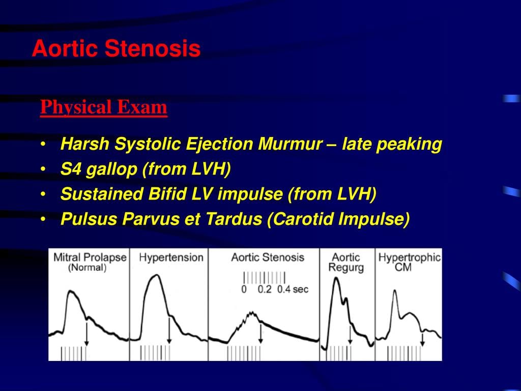
Pulsus parvus et tardus describes a pulse that is both weak (parvus) and late (tardus). A normal pulse feels strong and distinct with each heartbeat, but a pulsus parvus et tardus pulse is faint and delayed, as if struggling to emerge. This subtle difference, detectable by a trained healthcare professional, can be a critical indicator of an underlying cardiovascular issue, most commonly aortic stenosis.
The Aortic Stenosis Connection
The primary cause of pulsus parvus et tardus is usually aortic stenosis. The aortic valve, the exit door from your heart’s left ventricle to the aorta, narrows in this condition. This narrowing restricts blood flow, much like a kinked garden hose. The restricted flow weakens the pulse (parvus). The narrowed valve also slows the blood’s ejection, delaying the pulse (tardus).
While aortic stenosis is the most likely culprit, other conditions can also contribute to pulsus parvus et tardus. Severe heart failure, which weakens the heart muscle’s pumping ability, can also result in a similar weak and delayed pulse. Obstructions in the left ventricular outflow tract, the pathway from the left ventricle to the aorta, can similarly impede blood flow, mimicking the effects of aortic stenosis. To make sure you are purchasing the right product for your project, click on polyvinyl chloride conduit here.
Diagnosing Pulsus Parvus et Tardus and Aortic Stenosis
Doctors typically detect pulsus parvus et tardus during a routine physical exam by palpating the carotid artery in the neck. This initial assessment, while crucial, requires further investigation to confirm aortic stenosis and assess its severity. Tests like Doppler ultrasound and echocardiography provide detailed images of the heart’s structure and blood flow, helping determine the degree of valve narrowing.
Beyond the Pulse: Aortic Stenosis Symptoms
While pulsus parvus et tardus itself may not cause noticeable symptoms, the underlying aortic stenosis often does. These can include:
- Chest pain (angina): Reduced blood flow to the heart muscle causes this squeezing or tightness.
- Shortness of breath (dyspnea): The heart’s increased workload can lead to fluid buildup in the lungs, causing breathlessness, especially during exertion.
- Dizziness or fainting (syncope): Restricted blood flow to the brain can cause lightheadedness or fainting spells.
- Fatigue: The heart’s constant struggle to pump blood can lead to overall tiredness and weakness. Also, don’t miss our guide on protraction and retraction.
Treatment Approaches for Aortic Stenosis
The treatment for aortic stenosis depends on the severity. Lifestyle changes and medications may suffice for mild to moderate cases. These might include adopting a heart-healthy diet, engaging in regular exercise, and managing conditions like high blood pressure. Medications can help control symptoms like chest pain and hypertension.
Severe stenosis often requires more aggressive intervention, typically involving aortic valve replacement. This can be achieved through open-heart surgery or a less invasive procedure called transcatheter aortic valve replacement (TAVR). TAVR involves inserting a new valve through a catheter, typically through an artery in the leg or groin.
The Importance of Early Detection
Pulsus parvus et tardus, while a seemingly subtle sign, can be a valuable early warning for aortic stenosis. Early diagnosis and appropriate treatment are essential for effectively managing aortic stenosis and improving long-term heart health. Ongoing research continues to refine our understanding and treatment approaches, offering hope for improved outcomes for individuals with this condition.
Understanding the Mechanism of Pulsus Tardus
Pulsus tardus, the “delayed pulse” component of pulsus parvus et tardus, arises from the interplay of the narrowed aortic valve and the compliance (stretchiness) of the arterial walls downstream. The stenosis restricts blood flow, while the compliant vessel walls further dampen the pulse wave, delaying its arrival and diminishing its strength. This dampening effect on the pulse wave is what creates the characteristic “tardus” or late arrival of the pulse.
Hydrodynamic studies, using models to simulate blood flow, support this interaction between stenosis and vessel wall compliance as the primary mechanism behind pulsus tardus. The restricted and delayed blood flow also creates a distinct pattern on Doppler ultrasound known as the “tardus parvus” waveform. This waveform reflects the reduced blood flow magnitude during the heart’s contraction (systole).
Causes of Pulsus Parvus et Tardus
While aortic stenosis is the leading cause of pulsus parvus et tardus, other factors may contribute. These include:
- Left Ventricular Outflow Tract Obstruction: Any blockage in the pathway from the left ventricle to the aorta can restrict blood flow, similar to aortic stenosis.
- Severe Heart Failure: A weakened heart muscle struggles to pump blood efficiently, leading to a diminished and delayed pulse.
- Other less common conditions restricting blood flow: While rarer, other cardiovascular conditions can impact blood circulation, potentially contributing to pulsus parvus et tardus.
It’s crucial to remember that various conditions can restrict blood flow, and a thorough medical evaluation is essential to determine the precise cause.
Key Points About Pulsus Parvus et Tardus
- Definition: A weak (parvus) and delayed (tardus) pulse, a key clinical finding.
- Mechanism: Primarily caused by the combined effects of arterial stenosis (narrowing) and the compliance of the arterial wall downstream. This dampens the pulse, causing its delayed arrival.
- Primary Association: Strongly suggestive of aortic stenosis, a condition where the aortic valve narrows.
- Other Potential Causes: Includes left ventricular outflow tract obstruction, severe heart failure, and other conditions that restrict blood flow.
- Diagnosis: Detected through palpation of the arterial pulse and confirmed by Doppler ultrasound, which reveals a characteristic “tardus parvus” waveform.
- Symptoms: Often associated with symptoms of the underlying aortic stenosis, such as chest pain, shortness of breath, dizziness, and fatigue.
- Treatment: Focuses on managing the underlying cause, ranging from lifestyle changes and medications to surgical interventions like aortic valve replacement.
- Importance of Early Detection: Recognizing pulsus parvus et tardus is vital for timely intervention and the prevention of potential complications.
Disclaimer: This information is intended for educational purposes and does not substitute professional medical advice. Consult a healthcare professional for any concerns about your health.
- Photocell Sensors: A Complete Guide for Selection and Implementation - April 2, 2025
- Discover Famous French Women: A History of Impact - April 2, 2025
- 2025 World Map: Unveiling Geopolitical Shifts & Risks - April 2, 2025


