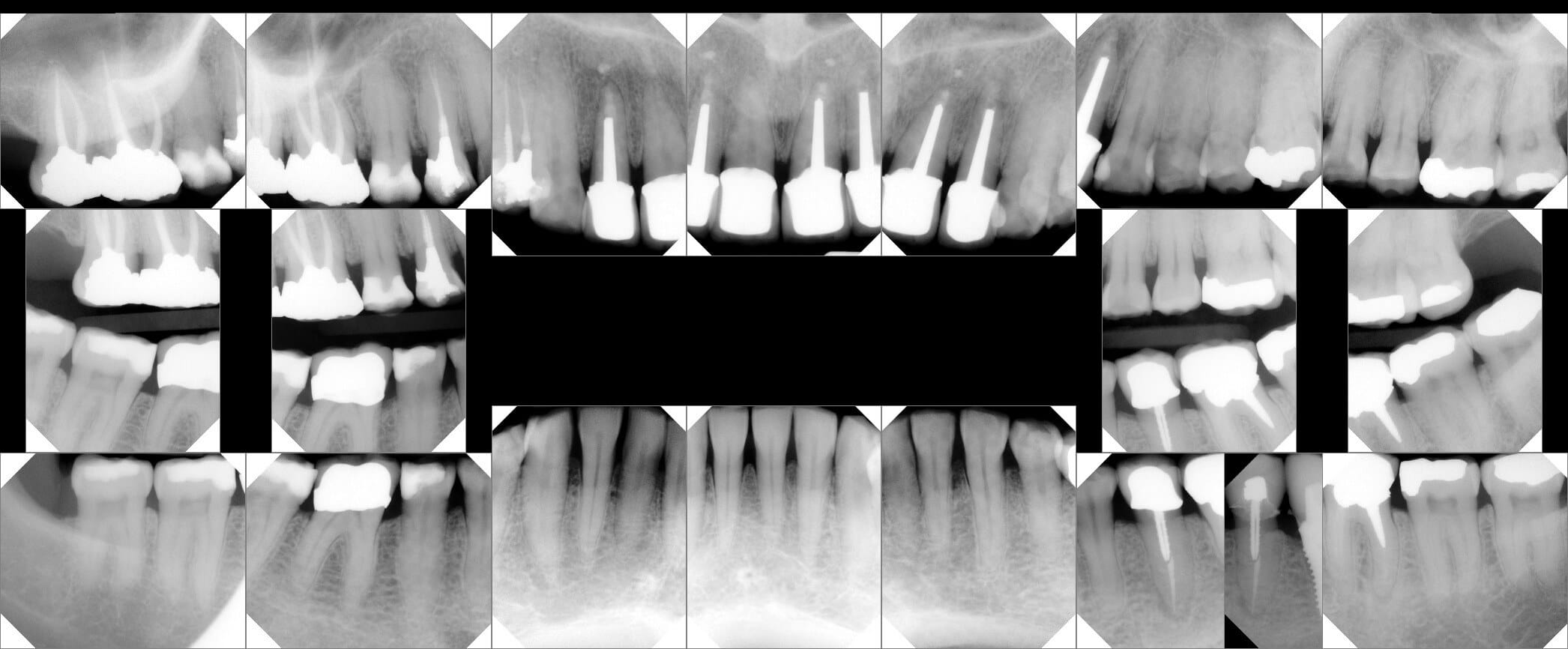
This guide provides a comprehensive overview of Full Mouth X-Rays (FMXs), empowering you to understand their role in maintaining optimal oral health.
Decoding the FMX
An FMX offers a detailed map of your teeth, jaws, and surrounding tissues, unveiling hidden issues often invisible to the naked eye. Think of it as a panoramic snapshot of your entire mouth, capturing everything from the crowns of your teeth to the roots and supporting bone structure. This comprehensive view enables dentists to diagnose a wide range of dental problems and develop personalized treatment plans.
What Makes Up an FMX?
A typical FMX comprises 18-20 individual radiographic images, each capturing a specific area of your mouth. These images can be broadly classified into two main types:
- Bitewing X-rays: These capture the crowns of your upper and lower teeth simultaneously, making them ideal for detecting cavities lurking between teeth and early signs of gum disease.
- Periapical X-rays: Providing a detailed view of the entire tooth, from crown to root, including the surrounding bone, periapical X-rays help identify infections, abscesses, and impacted teeth.
Why FMX X-Rays Matter
FMX X-rays offer a wealth of information crucial for maintaining a healthy smile:
Early Detection is Key
FMXs are incredibly effective at detecting hidden issues in their early stages. Tiny cavities, early signs of gum disease, and bone loss can be identified before they escalate into major problems, allowing for simpler and less invasive treatment.
The Big Picture of Oral Health
Like a panoramic photo, FMXs provide a comprehensive view of your entire mouth, not just isolated areas. This allows dentists to assess the overall health of your teeth, jawbone, and supporting structures, providing a more complete understanding of your oral health.
Precise Diagnosis for Effective Treatment
The clear images produced by FMXs enable accurate diagnoses, ensuring you receive the most appropriate and effective treatment. This precision is especially crucial for complex cases requiring specialized interventions.
Ensuring Patient Safety: Minimizing Radiation Risk
Modern digital X-ray technology used in FMXs significantly minimizes radiation exposure. While some researchers suggest potential long-term risks associated with even low levels of radiation, the current consensus among experts is that the benefits of early detection and accurate diagnosis far outweigh the minimal risks associated with dental X-rays. Dentists adhere to strict safety protocols, using protective aprons and thyroid collars to further minimize exposure. Ongoing research continues to refine safety practices and enhance our understanding of long-term effects.
What to Expect During an FMX
Getting an FMX is a quick, painless, and generally straightforward procedure. You’ll bite down on a small, comfortable sensor while the X-ray machine captures the images. The entire process usually takes just a few minutes.
FMX vs. CBCT: When is 3D Imaging Necessary?
While both FMXs and Cone Beam Computed Tomography (CBCT) scans provide detailed images, they differ in the level of information they offer. Think of an FMX as a 2D snapshot and a CBCT scan as a 3D movie of your mouth. CBCT provides highly detailed information about your facial bones and soft tissues, going beyond what traditional X-rays can reveal. CBCT is typically reserved for more complex cases, such as planning for dental implants, evaluating impacted teeth, or diagnosing temporomandibular joint (TMJ) disorders, where precise measurements and 3D visualization are crucial. Your dentist will determine which type of imaging best suits your individual needs. For example, a complex case like planning for dental implants may necessitate the use of CBCT for its detailed 3D imaging capabilities. Have you heard of the surgical procedure that treats certain pleural effusions? Click the link to know more about the technique of finger thoracostomy or the hepatorenal recess, between the liver and kidney that is very important in the development of certain hepatorenal syndromes.
Understanding Your Results: A Dialogue with Your Dentist
After the X-rays are taken, your dentist will carefully analyze the images, looking for any abnormalities. They will then discuss their findings with you, explaining what they see in clear, easy-to-understand terms and recommending any necessary treatment options. This is your chance to ask questions, address any concerns, and gain a deeper understanding of your oral health status.
FMX: A Vital Investment in Your Oral Health
FMX dental X-rays are fundamental to maintaining healthy teeth and gums. They provide a comprehensive view, enabling dentists to detect and address a wide range of dental issues, often before you experience any symptoms. While there might be slight variations in the specific X-rays taken, the overall goal is to provide your dentist with the clearest possible picture of your oral health. So, the next time you’re at the dentist, remember that FMXs are a valuable tool in your journey towards a healthier, happier smile. They offer invaluable insights that contribute to early detection, accurate diagnoses, and ultimately, a lifetime of good oral health. The cost of an FMX, typically ranging from $100 to $400 without insurance, can vary based on your location, insurance coverage, and the specific X-rays taken.
Diving Deeper into FMX Details
What Exactly is an FMX in Dental Terms?
An FMX provides a comprehensive view of your oral structures, surpassing what a routine visual examination can achieve. It’s a powerful tool encompassing all your teeth, from crowns to roots, the jawbone supporting them, and even adjacent soft tissues. This thorough examination helps dentists gain a complete understanding of your oral health.
The Components of an FMX
A standard FMX consists of 18-20 individual X-ray images, each taken from different angles to capture every aspect of your mouth. These images, like pieces of a puzzle, create a detailed picture of your oral landscape. Two main types of X-rays make up the FMX:
- Periapical X-rays: These focus on individual teeth, capturing their entire structure from crown to root tip and surrounding bone. They are particularly useful for detecting hidden issues like infections at the root tip or bone loss.
- Bitewing X-rays: Capturing the crowns of both your upper and lower teeth in a single shot, bitewing X-rays are essential for detecting cavities that often develop between teeth, areas often missed during visual exams.
The Importance of FMXs
FMXs play a vital role in three key areas of dental care:
- Uncovering Hidden Issues: Like a detective, FMXs help unearth hidden problems such as cavities between teeth, impacted wisdom teeth beneath the gums, lurking abscesses, and even bone loss or cysts. Early detection of these issues is paramount to preventing more serious problems later.
- Treatment Planning: Just as a blueprint is essential for house renovations, the comprehensive information from an FMX provides your dentist with the necessary insights to develop a personalized treatment plan addressing your specific needs. Whether it’s a simple filling or a more complex procedure, this complete picture leads to better treatment outcomes.
- Preventive Care: FMXs are crucial for preventative care. By identifying early signs of potential problems, they allow for timely intervention and preventive measures, akin to catching a small leak before it causes major damage. This proactive approach promotes long-term oral health.
FMX Safety and Ongoing Research
While radiation exposure is a valid concern, modern digital X-ray technology has significantly reduced the radiation used in FMXs. Dentists follow strict safety protocols using lead aprons and thyroid collars to further protect you during the procedure. While ongoing research explores potential long-term effects of even low levels of radiation, current expert consensus deems dental X-rays, including FMXs, safe, with the benefits outweighing the risks.
| Feature | Description |
|---|---|
| Number of Images | Typically comprises 18-20 individual x-rays. |
| Types of X-rays | Includes both periapical (individual tooth) and bitewing (upper and lower teeth together). |
| Purpose | Used for diagnosis of hidden issues, treatment planning, and preventive care. |
| Safety | Uses minimal radiation exposure with protective measures; generally considered safe, though research on long-term effects continues. |
In summary, an FMX serves as a detailed map of your mouth, guiding your dentist in delivering the best possible care. It facilitates the detection of hidden problems, effective treatment planning, and preventive measures to ensure a healthy smile. So, the next time your dentist recommends an FMX, remember it’s a powerful tool working to your advantage.
Unraveling FMX Codes in Dentistry
What are FMX Codes?
Beyond the procedure itself, understanding FMX dental codes is essential. These codes, officially known as “Intraoral comprehensive series of radiographic imaging codes”, are part of the Current Dental Terminology (CDT) system maintained by the American Dental Association (ADA). They are used to document and bill for FMX procedures, ensuring standardized record-keeping and insurance processing.
The Common Code: D0210
The most frequently encountered code is D0210, representing a complete set of intraoral X-rays of your teeth and surrounding bone. This code signifies a comprehensive evaluation of your oral structures.
Why are Full Mouth X-rays So Important?
FMXs are invaluable for uncovering hidden issues a regular checkup might miss, such as:
- Tiny cavities between teeth
- Early stages of gum disease
- Bone loss
- Impacted teeth
- Underlying infections
Early detection of these issues is critical for preventing larger, more complex, and costly problems down the line.
The FMX Process: A Quick and Efficient Overview
A typical FMX involves 14 to 22 individual images. A small sensor, placed inside your mouth, quickly captures these images digitally. Digital images offer superior clarity and detail compared to traditional X-rays, while also minimizing radiation exposure.
| What is it? | Explanation |
|---|---|
| FMX | Full Mouth X-ray – a comprehensive set of images of your entire mouth. |
| FMX Code | Special codes used to record and bill for these x-rays. |
| D0210 | The most frequently used code for a complete series of intraoral x-rays. |
| Why are they important? | To detect hidden dental issues like cavities, gum disease, and bone loss. |
| How many images? | Typically between 14 and 22, depending on the individual’s needs. |
Beyond D0210: Variations and Future Developments
While D0210 is the most common code, variations may exist depending on the specific types of X-rays taken or the equipment used. Ongoing research in dental imaging may lead to new codes and techniques in the future. Always consult your dentist if you have questions about the codes used for your X-rays. Remember, the exact number of x-rays in an FMX might vary slightly (14-22 is the typical range) based on individual dental anatomy and the dentist’s clinical judgment. This personalized approach ensures the most informative imaging for each patient.
Delving into the FMX Procedure
This section provides a detailed walkthrough of the FMX procedure, from start to finish.
What Happens During an FMX?
An FMX, or Full Mouth X-ray, is a comprehensive imaging technique that captures a panoramic snapshot of your entire mouth, including your teeth, jawbone, and surrounding soft tissues. This detailed view allows your dentist to gain a much clearer understanding of your overall oral health.
The FMX Process: A Step-by-Step Guide
Sensor Placement: You’ll bite down on a small sensor, similar to a tiny digital camera designed for your mouth.
Image Capture: This sensor captures multiple images from various angles, ensuring a thorough scan of every part of your mouth.
Digital Combination: The individual snapshots are then pieced together digitally, like a puzzle, to create a complete and detailed image of your mouth’s internal structure.
Why is an FMX Necessary?
The FMX is a powerful diagnostic tool. Like a detective searching for clues, it reveals hidden issues that might not be apparent during a routine check-up. This early detection is essential for effective treatment. For example, an FMX can detect:
Hidden Cavities: Cavities lurking between teeth, often undetectable during visual exams.
Early Gum Disease: Subtle signs of gum disease before noticeable symptoms appear.
Impacted Teeth: Teeth that haven’t fully erupted, potentially causing problems.
Cysts and Tumors: Abnormal growths within the mouth that require attention.
Bone Loss: Deterioration of the jawbone, a common issue associated with gum disease.
Types of FMX Procedures
There are several types of FMX procedures, and your dentist will choose the most appropriate based on your needs:
Digital FMX: The most common type, using a digital sensor for high-quality images with minimal radiation exposure.
Film-Based FMX: While less common now, traditional film-based FMXs are still used in some practices.
| FMX Type | Description | Benefits |
|---|---|---|
| Digital FMX | Uses a digital sensor to capture images. | Lower radiation, faster results, clearer images |
| Film-based FMX | Uses traditional x-ray film. | Still used in some practices, though less common now |
The FMX procedure is generally simple and painless, providing essential information about your oral health. It’s a valuable tool for developing a personalized treatment plan, keeping your smile healthy and bright. While experts often suggest regular FMXs, the ideal frequency depends on individual needs and risk factors. Discuss this with your dentist to determine the most suitable schedule for you. Ongoing research into x-ray technology and radiation exposure may refine best practices over time. Consult your dentist for the most up-to-date information.
- China II Review: Delicious Food & Speedy Service - April 17, 2025
- Understand Virginia’s Flag: History & Debate - April 17, 2025
- Explore Long Island’s Map: Unique Regions & Insights - April 17, 2025


1 thought on “FMX Dental X-Rays: A Comprehensive Guide to Understanding Your Oral Health”
Comments are closed.